
Fetal Pig Dissection Guide
Sample Pages
What follows are pages 52 and 53 of the Fetal Pig Dissection Guide. Note that each major structure is clearly described in the text, and that all descriptions are on the same page (52) as the illustration, or on the facing page (53).
Page 52
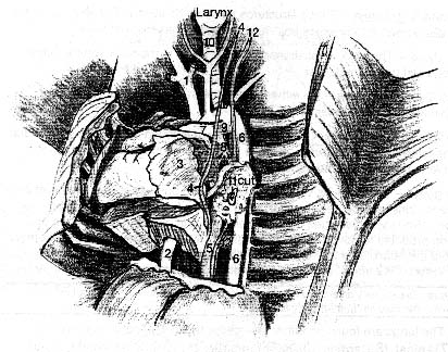
llustration of Chest Structures (smaller than actual size)
*Left lung removed for illustrative purposes.*
*Parts of the right lung are still visible.*
- Mediastinum — This is the space between the lungs. Organs in it include the heart, esophagus, aorta, and part of the thymus gland.
- 1) Anterior Vena Cava — In humans this is called the superior vena cava. This is a short vessel anterior to the right side of the heart. It carries blood to the right atrium.
- 2) Posterior Vena Cava — In humans this is called the inferior vena cava. This vessel returns blood from the posterior portion of the body. It can be viewed passing through the diaphragm mid-dorsally.
- 3) Pericardium — This is a membrane similar to the pleura. It also has a visceral part and a parietal part. This visceral pericardium is the thin, shiny membrane on the surface of the heart. The parietal pericardium is much thicker. (Peri = around. Cardio = heart.) There is a small amount of lubricating fluid found between the two layers, in a space known as the pericardial cavity.
Page 53
Medical Note
Inflammation of the pericardium is known as *pericarditis. Although it is painful and may be caused by a serious problem, pericarditis is not itself a life-threatening problem unless it is accompanied by fluid accumulation in the pericardial space. Such a fluid accumulation is known as *cardiac tamponade. It interferes with ventricular filling, reducing cardiac output, and can rapidly become life threatening.
- 4) Phrenic nerve — This nerve passes along the lateral edges of the pericardium. It supplies the muscle of the diaphragm.
- 5) Esophagus
- 6) Aorta — The large vessel you see first when looking at the front of the heart is the pulmonary trunk. The aorta is found behind the pulmonary trunk. The first part of the aorta, where it leaves the left ventricle is known as the ascending aorta. The curved part is known as the aortic arch. The rest of the aorta is known as the descending aorta. They look white because of their thick walls. If you have injected animals, they will have colored latex on the inside, but it is not visible externally.
- 7) Pulmonary Trunk — This artery leaves the heart on the ventral surface from the right ventricle and angles to the left.
- 8) Pulmonary arteries — The main pulmonary trunk divides into these vessels. In the adult, the pulmonary arteries carry deoxygenated blood to the lungs.
- 9) Ductus arteriosus — This short vessel in the fetal pig passes from the pulmonary artery to the aortic arch. Before birth it is used as a shunt to bypass the lungs, which are collapsed. In adults it becomes a small ligament. (See Fig. 43 – Fetal Circulation for a complete description of the fetal circulatory pattern.)
- Pulmonary veins — These 4 vessels can be traced returning from the lungs to the left atrium and should be injected with red dye (why should there be red dye in a vein?).
Trace the respiratory tract from larynx through the following structures:
- 10) Trachea — The structure below the larynx. Note its cartilagenous rings.
- 11) Bronchi — The branches of the trachea are known as bronchi. They also have cartilagenous rings. See Appendix 2, p. 100, for a more detailed description of bronchial structures.
Medical Note
An infection of the bronchi is known as #bronchitis. Usually many smaller subdivisions of the bronchi (the *bronchioles) are also involved in this inflammatory process.
- 12) Vagus nerve (*Cranial Nerve X) — the vagus nerve is somewhat dorsal to the phrenic nerve. On the left side of your pig (the pig’s left side), cut the diaphragm near where it attaches to the chest wall, laterally and dorsally. Gently lift up the lungs as far as you can. You will see several prominent structures.
Fetal Pig Dissection Guide
113 pages, 63 illustrations, 33 medical notes. Coil bound.
Last updated Sept., 2004
Learn more about the Biology major at Goshen College.